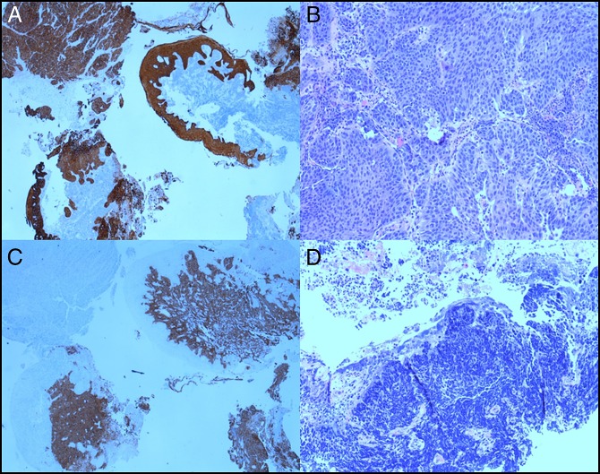
Figure 2.
Squamous cell carcinoma of the esophagus demonstrating (A) atypical cells with positive staining for cytokeratin 5/6 immunostain; (B) squamous differentiation with increased mitosis (hematoxylin and eosin stain, 10×); (C) positive staining for sign apt to 57 within the same tumor confirms neuroendocrine differentiation; and (D) the neuroendocrine tumor demonstrates atypical cells with hyperchromatic nuclei and increased nuclear/cytoplasmic (hematoxylin and eosin stain, 10×).

