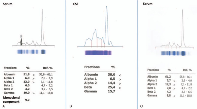
Figure 1.

(A) Serum electrophoresis before treatment initiation demonstrating a monoclonal M-paraprotein of 0.61 g/dL. (B) Cerebrospinal fluid (CSF) electrophoresis showing a monoclonal component in the gamma region; immunofixation revealed immunoglobulin M and kappa light-chain monoclonal bands. (C) Serum electrophoresis after 6 months of treatment initiation showing disappearance of the monoclonal band, whereas serum immunofixation was negative.
