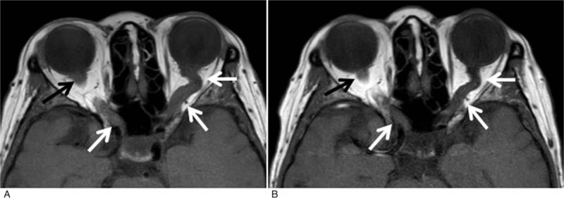
Figure 2.

(A) At presentation brain magnetic resonance imaging revealed bilateral swelling of optic nerves extending from the retina to the optic chiasm and swelling of the left optic tract. T1-weighted image in the axial plane shows diffuse enlargement of both optic nerves (arrows). (B) Corresponding T1-weighted image in the axial plane of the same patient 3 months after treatment demonstrates decrease in the size of both nerves.
