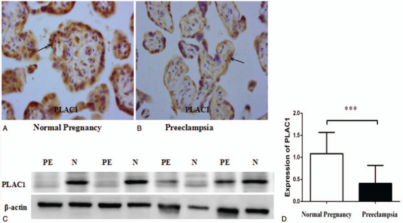
Figure 1.

Immunohistochemistry staining for PLAC1 and expression of PLAC1 protein in placenta between normal pregnancy (N) and severe preeclampsia (PE). The positive brown staining in the placental tissues show that PLAC1 expressed in the trophoblast both normal pregnancy (A) and severe preeclampsia (B). PLAC1 protein (30 ug per lane) extracts from placental tissues of normal pregnancy and severe preeclampsia was determined by Western blotting, and there were significant decrease of PLAC1 expression in severe preeclampsia (PE) than control (C, D). Values represent mean ± SD, n = 19 per group, ∗∗∗P < .001.
