
Figure 2.
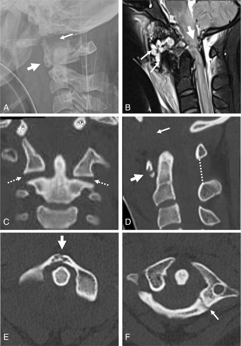
Lateral radiograph of the cervical spine revealed increased soft tissue swelling, horizontal split fracture of the anterior arch of the atlas (thick arrow), and increased basion-dental interval of 24 mm (arrow) (A). Sagittal MRI revealed complete rupture of the ligamentous complex (white arrows) and severe intramedullary cord hemorrhage at the occipito-cervical junction (thick arrow) (B). Coronal 2-D CT revealed vertical displacement of the occipital condyles (asterisks), and increased atlanto-axial joints (dotted arrows) (C). Sagittal 2-D CT revealed horizontal split fracture of the anterior arch of the atlas (thick arrows), increased atlanto-axial joints (dotted arrows), and increased basion-dental interval of 24 mm (arrow) (D). Axial CT revealed 2-part fracture of the atlas, including anterior arch fracture (thick arrow) and posterior arch fracture (arrow) (E and F). Parasagittal 2-D CT scans demonstrated vertical displacement of the occipital condyles (asterisks) with respect to the lateral mass of the atlas and posterior arch fracture of the atlas (arrowhead) (G and H). At 1 year post-surgery, plain radiographs of the cervical spine revealed solid fusion of the occipitocervical junction (I and J).