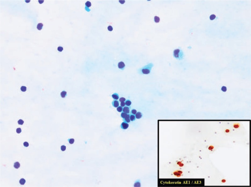
Figure 1.

Cerebrospinal fluid with an infiltration by ductal breast carcinoma. Isolated cells and poorly cohesive cluster of cells. Eccentric nuclei often protruding from the cytoplasm. Enlarged, variably hyperchromatic nuclei in a clean background. In the image in the lower right corner, we can see positive immunoreaction for Cytokeratin AE1/AE3. This is concordant with an infiltration by carcinoma. Alvaro Gutierrez Domingo, MD, Pathological Department, Virgen Macarena Hospital, Sevilla (Spain).
