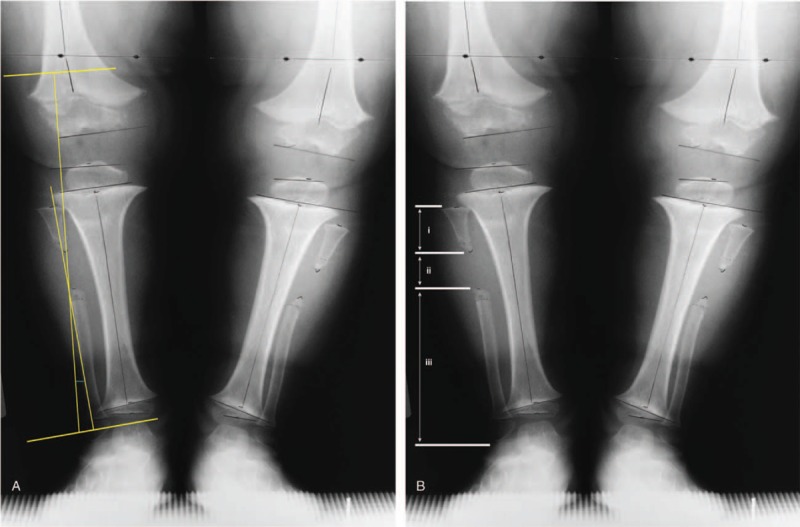
Figure 1.

Description of the following radiographic measurements taken: (A) tibia varus and (B) location of the fibular excision. Tibia varus was measured similar to the Cobb angle, where a line was drawn parallel to the tibial plafond and a second line drawn parallel to the proximal tibial condyles and the angle formed by the intersection of the lines perpendicular to these parallel lines representing tibia varus or valgus. The fibular excision site was calculated using the following measures: (i) length between the head of the fibula and first cut; (ii) the gap created by the excision, and (iii) length from the distal cut to the lateral malleolus. A ratio (i/[i + ii + iii]) with a value less than 0.30 was considered a proximal excision and a value greater than or equal to 0.30 was considered a mid-shaft incision.
