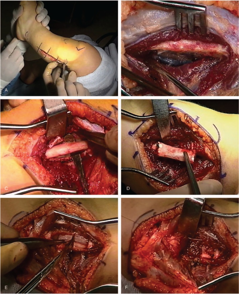
Figure 2.

Images from surgical procedure depicting the following: (A) skin incision; (B) exposure of fibula with periosteum intact; (C) sub-periosteal exposure of the fibula; (D) excision of fibula; (E) splitting of the periosteum, and; (F) suturing of periosteum and muscle fibers over bone end.
