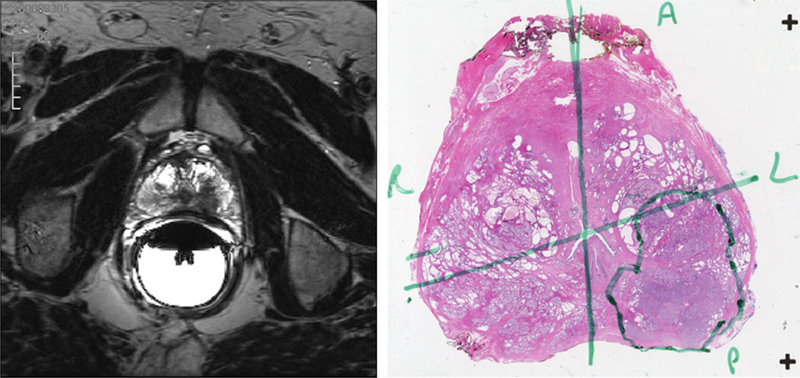
Fig. 1.
One example prostate tumor from the dataset used in this study. A single axial slice of the prostate, as visualized on high-resolution 3D axial TSE (turbo spin echo) T2-weighted MR (left) and the closest matching whole-mount section from histopathology (right). The tumor was localized on the left posterior region of the prostate as seen by the low MR signal intensity areas in the otherwise high MR signal intensity peripheral zone. The MR findings were confirmed through histopathology; the tumor as localized by the pathologist is annotated in green.

