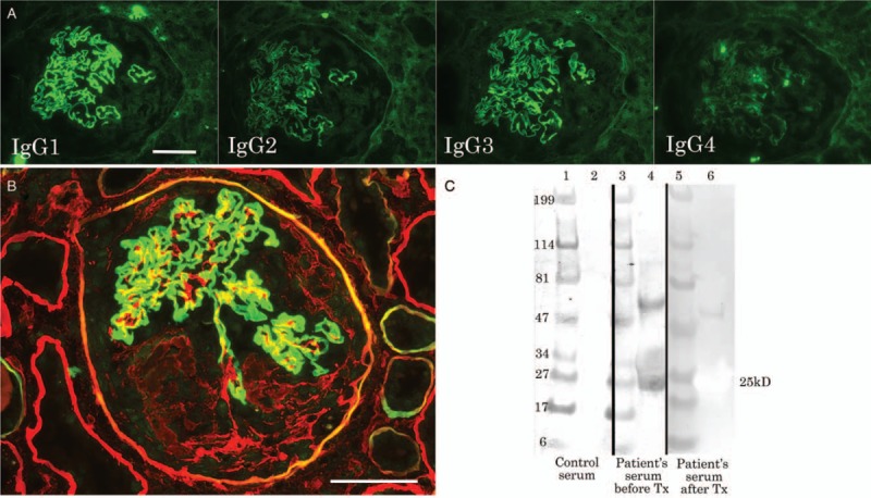
Figure 2.

Immunofluorescence for IgG subclass (A), double staining for type IV collagen α2 (red) and α5 (green) (B) and immunoblotting for NC1 domain protein of type IV collagen α3 (lane 2, 4, 6) and molecular weight markers (lane 1, 3, 5) with the patient's serum before (3, 4) and after (5, 6) treatment as well as control serum (1, 2) (C). The bars indicate 50 μm.
