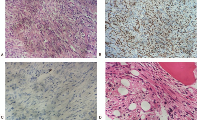
Figure 2.

(A) Histological examination demonstrated that lung lesion was composed of spindle cells in the background of plasma cells and lymphocytes (hematoxylin and eosin stain, ×400). (B) Immunohistochemistry analysis showed that the spindle cells were positive for smooth muscle actin (×200). (C) Immunohistochemistry analysis showed that the spindle cells were negative for anaplastic lymphoma kinase 1 (×400). (D) Bone marrow was infiltrated by spindle cells (hematoxylin and eosin stain, ×400).
