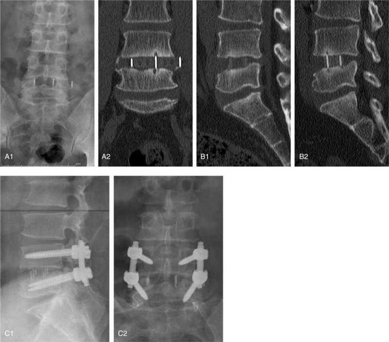
Figure 1.

A 34-year-old male patient underwent XLIF 3 months ago, and allogeneic bone was used as bone filling material during the operation. At 3 months after operation X-ray (A1) and coronal position of CT plain scan (A2) showed that cage displacement was obvious, sagittal position of CT plain scan (B1, B2) indicated that there was no osseous connection between the implant and the upper and lower endplates. The patients underwent routine posterior surgery again (C1, C2). CT = computed tomography, XLIF = extreme lateral interbody fusion.
