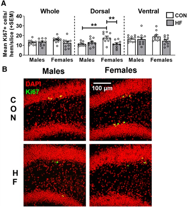
Figure 2.
A, Cell proliferation in the dentate gyrus, as measured by the mean (±SEM) number of Ki67+ cells per hemisphere per 40 μm slice throughout the whole hippocampus, and in the dorsal and ventral subregions. On a control diet, females had a greater number of Ki67+ cells in the dorsal hippocampus compared with males, while HF diet reduced the number of Ki67+ cells in the dorsal hippocampus of females only. N = 10/group. **p < 0.01. B, Representative images of Ki67 (green) and DAPI (red) immunostaining in the dorsal hippocampus. Estimation statistics for all neurogenesis-related measures can be seen in Extended Data Figure 2-1.

