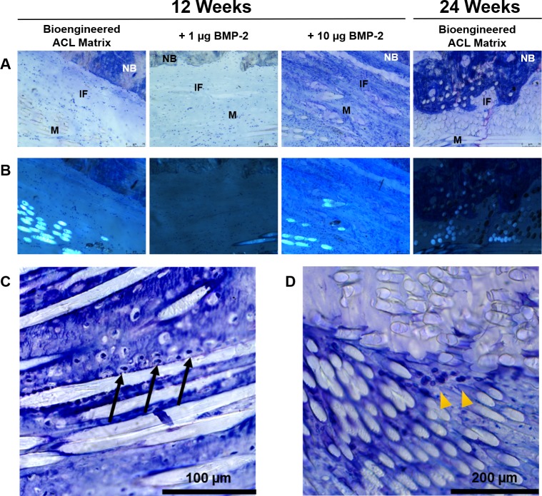
Fig 6. Toluidine Blue stain of the femoral tunnel matrix-to-bone interface.
A) 80x images in the mid-center of the bone tunnel region. Minimal proteoglycan was observed at the matrix-to-bone interface in all 12-week samples (proteoglycans stain purple). At 24-weeks, the bioengineered ACL matrix was incorporated with mature bone. B) Polarized images of the same region in panel A. C) Toluidine Blue staining of the aperture of the femoral bone tunnel at 12-weeks. Proteoglycan content was observed (purple staining) and accompanied by the presence of chondrocyte-like cells (black arrows). D) Presence of chondrocyte-like cells (yellow arrowheads) found in the center of the bioengineered ACL matrix. NB, native bone; IF, interface; M, matrix.

