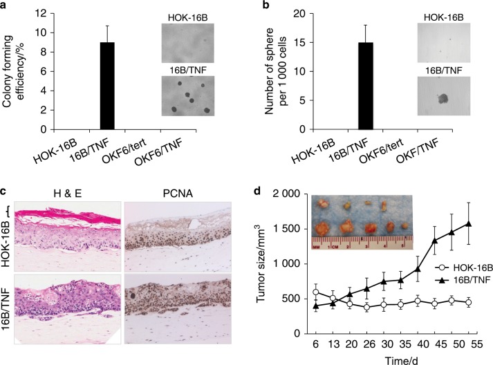
Fig. 2. Chronic TNFα exposure induces malignant growth properties in HPV-immortalized oral keratinocytes.
a Anchorage-independent growth ability was determined by soft agar assay. Five thousand cells were seeded on 0.4% soft agar and incubated for 3 weeks. Colonies were counted, and images were acquired at a magnification of 40×. The assay was performed in the absence of TNFα. b Self-renewal capacity was determined by tumor sphere formation assay. Single cells were plated in ultralow attachment plates at a density of 1 000 cells per mL in serum-free tumor sphere-forming medium. Tumor spheres were counted on day 17, and images were acquired at a magnification of ×40. The organotypic raft assay was performed in the absence of TNFα. c Organotypic raft cultures were established with HOK-16B and 16B/TNF cells. After 14 days of air lifting, the mucosal tissue constructs were harvested and processed for H&E staining and immunohistochemical staining against PCNA. Bracket indicates a terminally differentiated cornified layer, which is missing in the raft culture of 16B/TNF cells. Slides were scanned at ×40 magnification. The organotypic raft assay was performed in the absence of TNFα. d In vivo tumourigenicity was determined by xenograft tumor assay. HOK-16B and 16B/TNF cells were injected subcutaneously into five nude mice.

