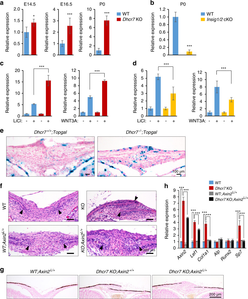
Fig. 6.
Altered WNT/β-catenin signaling in calvaria from Dhcr7 and Insig1/2 mutant mice during craniofacial development. a Quantitative RT-PCR for Axin2 in calvaria from wild-type (WT; blue bars) control and Dhcr7 knockout (KO; red bars) mice at E14.5, E16.5, and P0 (newborn). n = 6 per group. *P < 0.05; ***p < 0.001. b Quantitative RT-PCR for Axin2 in calvaria from newborn WT (blue bar) and Insig1/2 conditional KO (cKO; yellow bar) mice. n = 6 per group. ***P < 0.001. c Quantitative RT-PCR for Axin2 after treatment with LiCl (left panel) or WNT3A (right panel) in WT (blue bars) and Dhcr7 KO (red bars) osteoblasts. n = 6 per group. ***P < 0.001. d Quantitative RT-PCR for Axin2 after treatment with LiCl (left panel) or WNT3A (right panel) in WT (blue bars) and Insig1/2 cKO (yellow bars) osteoblasts. n = 6 per group. ***P < 0.001. e β-galactosidase staining (blue) for sites of WNT/β-catenin signaling activation in the frontal bones of P0 Dhcr7+/+;Topgal and Dhcr7−/−;Topgal mice. Nuclei were stained with nuclear fast red. Scale bars, 100 µm. f Hematoxylin and Eosin staining of the sagittal sutures of newborn WT, WT;Axin2L/+, Dhcr7 KO and Dhcr7 KO;Axin2L/+ mice. Arrowheads indicate the osteogenic front. Accelerated bone formation of the sutures (frontal, coronal, and sagittal sutures) was normalized in newborn Dhcr7 KO;Axin2L/+ mice (n = 6/6). Scale bars, 100 µm. g Von Kossa staining of the sagittal sutures of newborn WT;Axin2L/+, Dhcr7 KO and Dhcr7 KO;Axin2L/+ mice. Scale bar, 200 µm. h Quantitative RT-PCR of the indicated genes in newborn WT (blue bars), Dhcr7 KO (red bars), WT;Axin2L/+ (gray bars) and Dhcr7KO;Axin2L/+ (black bars) mice. n = 6 per group. ***P < 0.001.

