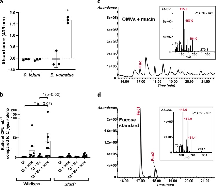
Fig. 1. C. jejuni and B. vulgatus use of l-fucose and mucin degradation.
a Fucosidase activity detected by cleavage of 4-nitrophenyl-α-l-fucopyranoside measured by absorbance at 405 nm after 18 h incubation with the substrate under appropriate bacterial growth conditions (gray bars are at t = 0 h and white bars are at t = 18 h). Values represent means of three technical replicates and error bars show one standard deviation. Values from each replicate are overlaid as dots. The asterisk indicates a significant increase in absorbance (p = 0.02) compared to t = 0. b Ratios of C. jejuni (Cj) wildtype and fucP mutant in co-culture with B. vulgatus (Bv) and mucin (Muc) in minimal essential medium α. Values of bars represent means from eight biological replicates and error bars show one standard of the mean. Values from each replicate are overlaid as dots. Asterisks denote significance with p-values denoted in parentheses as determined by two-way ANOVA with Tukey post-test. c GC-MS chromatogram and spectrum (inset) of l-fucose released from mucin treated with B. vulgatus outer membrane vesicles (OMVs). d GC-MS chromatogram and spectrum (inset) of the l-fucose standard used to confirm the identity of l-fucose in the OMV-treated mucin sample. Fragment ions (Fuc1 and 2) are highlighted in red and exist in both α- and β- configurations and correspond to expected fragmentation patterns of fucose illustrated in Supplementary Fig. 1b.

