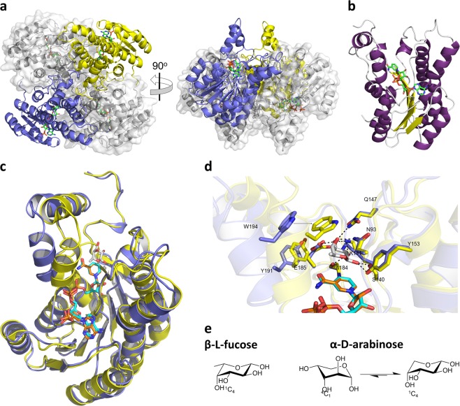
Fig. 2. FucX crystal structure.
a The tetrameric organization of FucX observed in complex with NADP+. Two monomers are shown in blue and yellow cartoon representation and the other two in grey with transparent solvent accessible surfaces. Bound NADP+ is shown as green sticks. b Cartoon representation of the FucX monomer with β-strands shown in yellow, α-helices shown in purple, and loops shown in gray. Bound NADP+ is shown as green sticks. c Structural alignment of C. jejuni FucX (in blue) in complex with NADP+ (in cyan) with the homolog from Burkholderia multivorans (4GVX, in yellow) in complex with NADP+ (in orange) and l-fucose (in gray). d Close-up of the active site alignment of FucX (in blue) in complex with NADP+ (in cyan) with the homolog from B. multivorans (4GVX, in yellow) in complex with NADP+ (in orange) and L-fucose (in gray). Dashed lines indicate hydrogen bonds. E) Chair conformations of α-l-fucose and β-d-arabinose.

