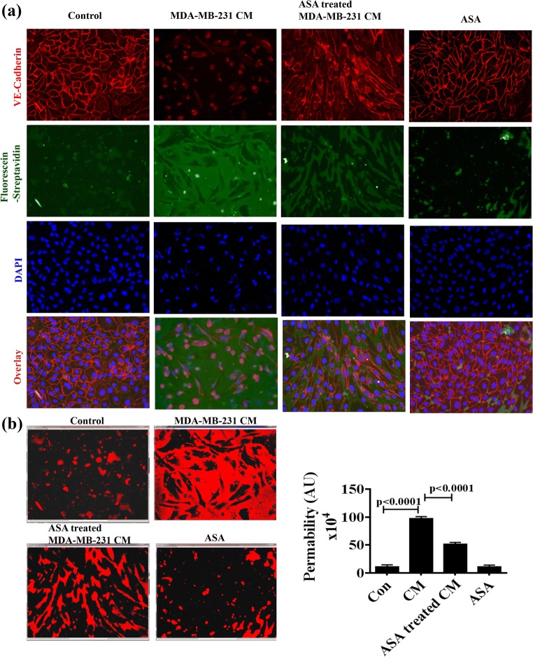
Fig. 5.
Effects of ASA treated or untreated MDA-MB-231-CM on vascular permeability in vitro. a HUVEC monolayer was formed on biotinylated gelatin on 8-well chamber slides as described in methods. Cell were treated with regular medium, untreated or ASA pretreated MDA-MB-231-CM or ASA alone for 48 h. Then Fluorescein-streptavidin was added as indicated in the protocol. Fixation and staining with anti-VE-cadherin (red) and DAPI (blue) were performed. Green fluorescence imaging between cell-cell junction was considered as permeable area and immunofluorescence staining of VE-cadherin (Red) detect cellular junctional integrity. b Permeabilization was quantified by of FITC coupled fluorescein-streptavidin using image J software. The histogram shows the positive staining of fluorescein-streptavidin and the bar diagram represents the quantitation of permeability. Photograph was taken under fluorescence microscope with magnification of X250. Data shows mean ± SD and represents at least 3 independent experiments. For statistical analysis two paired student’s t test was done

