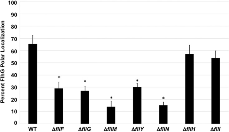
FIG 8.

FlhG polar localization in WT C. jejuni and isogenic flagellar mutants. Strains were analyzed by immunofluorescent microscopy after staining with both FlhG and whole C. jejuni antisera. Cells in which detection of FlhG was exclusively at poles were considered positive for polar localization of FlhG. Each strain was analyzed in triplicate, and at least 100 individual cells were counted per sample. After analysis, the percentage of cells with exclusive polar localization of FlhG were averaged, and the standard deviations were determined (bars). A Student's t test was performed to determine the statistical significance of differences in polar localization of FlhG between WT and mutant strains (*, P < 0.05).
