Figure 3:
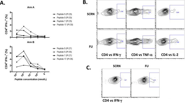
Recognition of processed NY-ESO-1 protein by vaccine-induced NY-ESO-1-specific T cells. A. Avidity of vaccine-induced NY-ESO-1-specific CD4+ T cells, after vaccination (FU) is shown. PBMCs from patients in arm A and arm B (2 from each group) were cultured in the presence of NY-ESO-1 OLPs for 12 days. On that day, cells were re-stimulated with serial dilutions of NY-ESO-1-specific peptides (1 μM-1 nM) (P5, 7, and/or 17) and GolgiPlug as well as GolgiSTOP were added after 1h of peptide stimulation. Then, intracellular cytokine staining was performed to assess flow cytometry. Percentage of CD4+ IFN-γ+ cells were determined at different peptide concentrations; peptides: P5 (AA81–100), P7 (AA119–143), and P17 (AA161–180). N=2 (arm A; patient 25 responded against P5 and 7; patient 33 responded against P5 and 17), n=2 (arm B; patient 27 responded against P5 and 7; patient 35 responded against P5 and 17). B. Recognition of NY-ESO-1 peptides (OLPs) presented by MoDCs. MoDCs from an arm B patient (patient 32) were pulsed with NY-ESO-1 OLPs for 4 h and then co-cultured with PBMCs isolated from screening and FU time points, or C. PBMCs from the same patient were expanded for 10 days in the presence of NY-ESO-1 OLPs. At day 10, cells were harvested and re-stimulated with MoDCs pulsed with OLPs (1 μM) and GolgiPlug as well as GolgiSTOP were added after 1h of peptide stimulation. Intracellular cytokine staining was performed to assess flow cytometry.

