Figure 2.
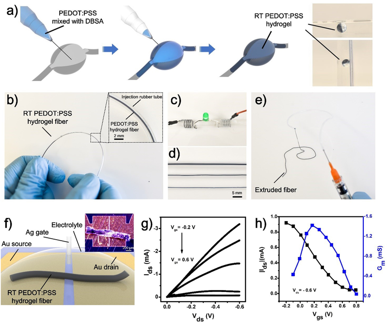
a) Schematic of injectable RT-PEDOT:PSS hydrogels: Puncturing the soft tissue with syringe; Syringe-injecting PEDOT:PSS suspension (4 v/v.% DBSA) into the cavity; and RT-PEDOT:PSS hydrogel formed spontaneously after 10 min; The optical images show the RT-PEDOT:PSS hydrogel attached to the wall of the cavity even we tilted the mold; b) Formation of the RT-PEDOT:PSS hydrogel in a plastic tube via syringe injection. Inset: enlarged view of the RT-PEDOT:PSS hydrogel in tube; c) Application of RT-PEDOT:PSS hydrogel fibers for driving an LED; d) Injectable RT-PEDOT:PSS hydrogel fibers with different diameters of 875, 480, and 400 μm; e) Extruded RT-PEDOT:PSS hydrogel fibers with a syringe; f) Schematic of the fabricated OECTs with injected RT-PEDOT:PSS hydrogel fiber. The hydrogel fiber was freeze-dried after printing on the Au electrodes which have a gap of 10 μm; the inset shows the real optical image of the freeze-dried fiber on the source-drain electrodes. g-h) Output and transfer curves of the OECTs with RT-PEDOT:PSS hydrogel fibers as the channel.

