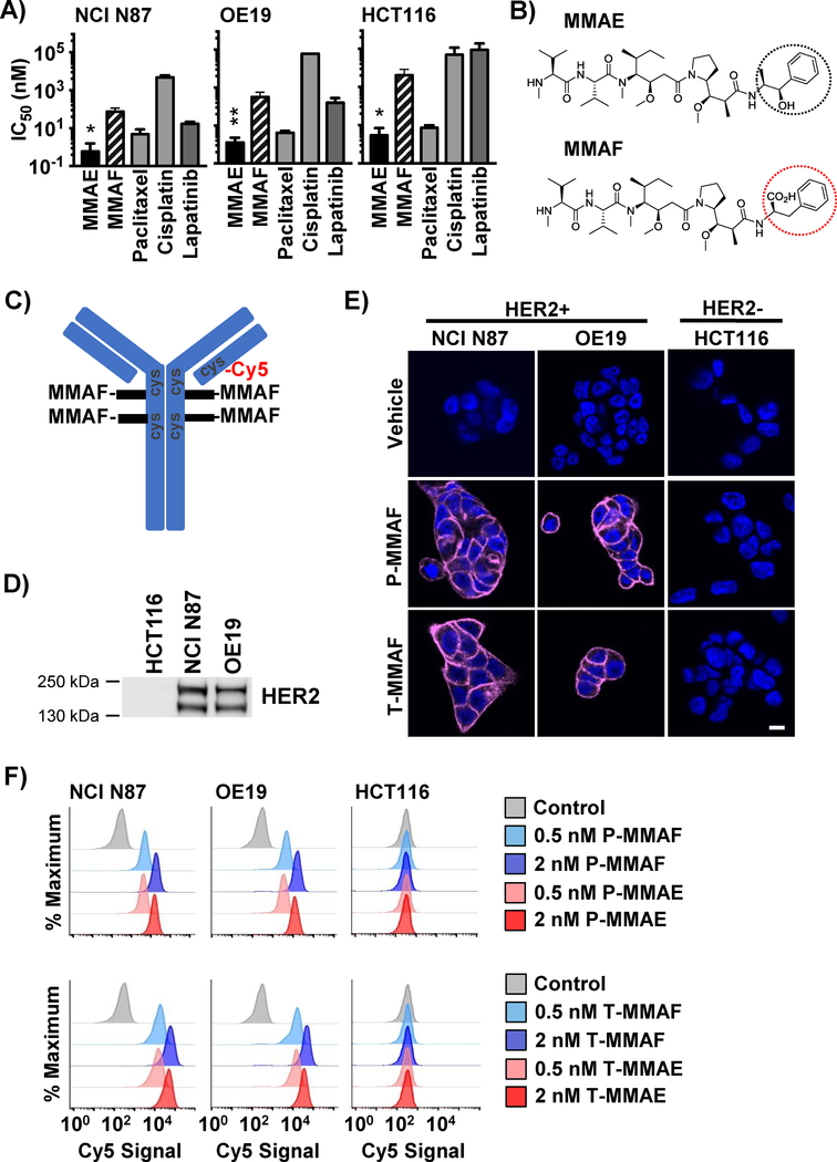
Figure 1: MMAF antibody conjugates selectively bind HER2 expressing cells.
A) IC50 of monomethyl auristatin E (MMAE) and MMAF compared to chemotherapy drugs in NCI N87, OE19 and HCT116 cells. B) Chemical structures of MMAE and MMAF with differences circled. C) Schematic representation of 4 MMAF drugs attached to a targeting antibody through maleimidocaproyl-valine citrulline-para amino benzyl carbamoyl linkers and Cy5 attached through maleimide to reduced hinge disulfides of cysteine. D) HER2 expression in cell lines. Immunoblot for total HER2 from cell lysates. E) ADC binding to HER2 positive (NCI N87 and OE19) and HER2 negative (HCT116) cells by microscopy. Cells exposed to 2 nM of Cy5 labeled MMAF conjugated to pertuzumab (P-MMAF) or trastuzumab (T-MMAF) for 30 minutes and then incubated in drug free media. Cells fixed 2 hrs later and imaged for Cy5 fluorescence (magenta). Nuclei stained with DAPI (blue). White scale bar, 10 μm. F) Cell surface binding of ADC by flow cytometry. Cells incubated on ice with Cy5 labeled ADC and Cy5 signal measured by flow cytometry and plotted as percent of maximum. Each peak normalized to the peak height at its mode of distribution resulting in each peak’s maximum set at 100%. *P<0.05, **P<0.01.

