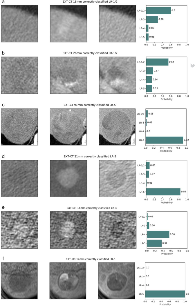
Fig. 5.
Examples of correctly categorized (LI-RADS v2014) observations on external datasets: (a–d) EXT-CT and (e, f) EXT-MR. Each plot title includes the dataset name, observation diameter, ground truth, and predicted LI-RADS category. Pre-contrast, late arterial, and delayed phase cropped images are displayed from left to right, and the rightmost bar plot shows the output probability for each LI-RADS category. (a) transient arterial phase hyperenhancement (LR-1/2), (b) typical hemangioma (LR-1/2), (c, d, f) HCC (LR-5), (e) probable HCC (LR-4).
Abbreviations: HCC, hepatocellular carcinoma, LI-RADS and LR-, the Liver Imaging Reporting and Data System

