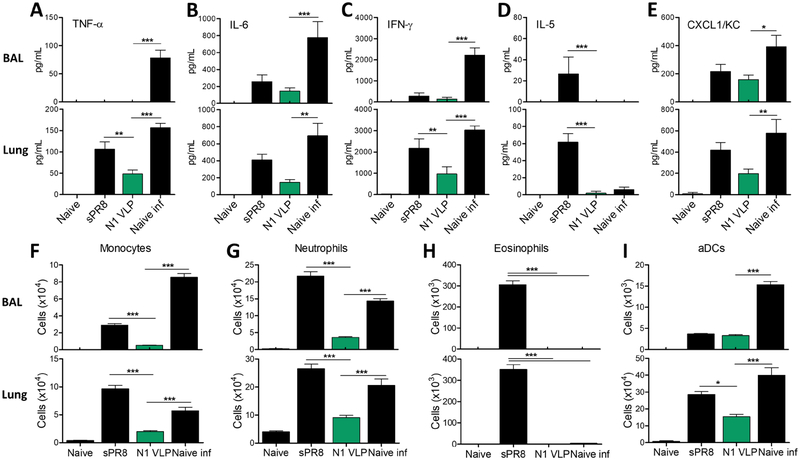
Figure 5. N1 VLP vaccination prevents lung inflammation due to heterologous rgH5N1 virus infection.
Inflammatory cytokines, chemokines, and cellular phenotypes were determined in BALF and lung samples collected at 7 dpi with rgH5N1 virus. (A-E) ELISA of cytokines and chemokines in BALF and lungs. (A) TNF-α. (B) IL-6. (C) IFN-γ. (D) IL-5. (E) chemokine CXCL/KC. (F-I) Phenotypes of cellular infiltrates as determined by flow cytometry. (F) Monocytes (CD11b+Ly6chiF4/80+). (G) Neutrophils (CD11b+Ly6c+F4/80−). (H) Eosinophils (CD11b+CD11c+SiglecF+). (I) Activated dendritic cells (aDCs, CD45+CD11b+MHCII+). Naïve: unvaccinated mice without virus infection. sPR8: split sPR8 vaccinated mice with rgH5N1 virus infection. N1 VLP: N1 VLP vaccinated mice with rgH5N1 virus infection. Naïve inf: unvaccinated mice with rgH5N1 virus infection. Statistical significance was determined by using one-way and dunnett’s multiple comparison test ANOVA. Data (n=4) are representative of individual animal out of two independent experiments. Error bars indicate the means ± SEM. *, p<0.05, **, p<0.01, ***, p<0.001.

