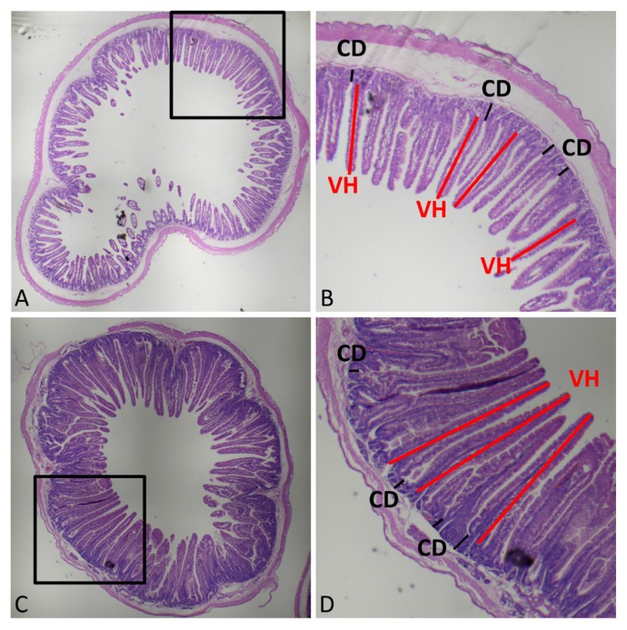
Figure 1.
Histomorphological evaluation of ileal and jejunal tissues. Morphological evaluation of small intestinal villi was performed with hematoxylin and eosin (H&E) staining. (A) and (C) represent morphology of the ileum and jejunum, respectively. (B) and (D) represent the enlarged areas of (A) and (C), respectively. VH, villus height; CD, crypt depth. Three fields of fluff integrity and straightness with 10 complete fluffs (length of VH and CD) were selected randomly and measured in each slice.

