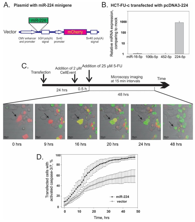
Figure 2.
Overexpression of miR-224-5p promotes the apoptosis of HCT-FU-c cells after treatment by 5-fluorouracil. (A). A schematic presentation of the cassette for the expression of miR-224 minigene and red fluorescence protein mCherry inserted within the pcDNA3 vector. The miR-224 coding minigene and the reporter gene are placed downstream of CMV and the SV40 promoter, respectively. (B). miR-16-5p, miR-106b-5p, miR-452-5p and miR-224-5p relative expression after transfection of HCT-FU-c cells with pcDNA3-224 plasmid comparing with HCT-FU-c cell transfected with vector. RNA was isolated after 24 h. (C). Scheme of caspase 3 and 7 activity analysis experiment and representative microscopy images. Cells were transfected 24 h prior to addition of CellEventTM dye. Then, 30 min after transfected cells were treated with 25 µM 5-FU, the fluorescence of cells was imaged at 15 min intervals for 48 h. Representative microscopy images of the sample captured at different time points. Red signal indicates transfected cells expressing mCherry; green—cells with activated caspase 3/7; yellow—transfected cells with activated caspase 3/7. (D). Real-time kinetics of caspase 3/7 activation in cells transfected with miR-224 overexpression plasmid (black dots) or vector (grey dots) in response to 5-FU treatment. The sum effect of the activated caspases is presented as the average ± SD of three biological experiments.

