Figure 6.
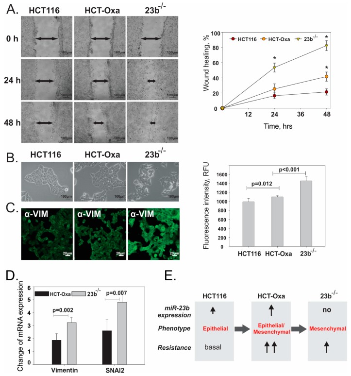
Evaluation of the migration potential and EMT status of HCT116, HCT-Oxa-c and 23b−/− cells. (A). Estimation of cell migration capabilities using wound healing assay. (B). Identification of cell morphology by bright field microscopy. (C). Assessment of mesenchymal marker vimentin by confocal microscopy. (D). Analysis of mesenchymal cell marker vimentin and EMT transcription factor SNAI2 by qPCR. (E). Schematic representation of EMT status and resistance of HCT116, HCT-Oxa-c and 23b−/− cells, showing the importance of partial EMT for cancer cell resistance.

