
Figure 2.
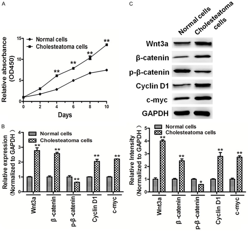
Cholesteatoma cells showed high cell growth activity and induced Wnt/β-catenin signaling. A. Cells were seeded and cultured in 96-well plates. Cell viability was assayed using a CCK8 kit according to the manufacturer’s instruction at indicated time points. The results showed that cholesteatoma cells showed high cell growth activity compared with normal cells. B, C. qRT-PCR and western blotting showed that cholesteatoma cells induced Wnt/β-catenin signaling, the expression levels of β-catenin, Cyclin D1 and c-myc were up-regulated in cholesteatoma cells, and the expression levels of p-β-catenin was down-regulated in cholesteatoma cells. The density of protein levels of above was quantified by ImageJ software and normalized to the level of GAPDH. Data represent mean ± SD of three replicates. *Indicates significant difference at P<0.05. **Indicates significant difference at P<0.01.