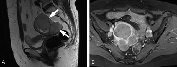
Figure 1.

MRI features of large cell neuroendocrine carcinoma of the endometrium in a 61-year-old woman. A. Sagittal T2-weighted images showing uterine and parauterine masses (arrows) of intermediate signal intensity. B. Contrast-enhanced fat-suppressed T1-weighted image showing mass enhancements.
