
Figure 2.
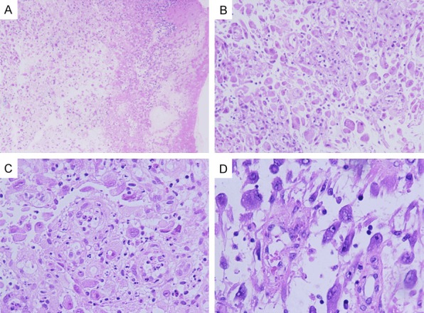
Microscopic features of stomach EIMS. A. Marked necrosis (magnification, × 50). B. Tumor cells were predominantly composed of non-cohesive sheets or clusters of round to epithelioid cells and embedded in a loose or myxoid stroma, with abundant neutrophilic granulocyte (magnification, × 100). C. Epithelioid cells were characterized by abundant eosinophilias cytoplasm, obvious vesicular nuclei with prominent nucleoli (magnification, × 200). D. Ganglion cell-like cells with abundant basophilic cytoplasm, obvious vesicular nuclei and prominent nucleoli (magnification, × 200).