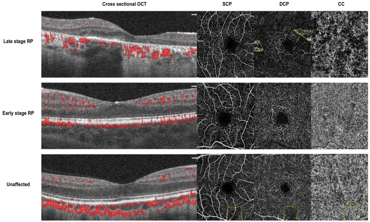
Figure 1.
Cross sectional optical coherence tomography (OCT) with angio flow (denoted in red) and 3 × 3mm en face optical coherence tomography angiography (OCTA) of the superficial capillary plexus (SCP), deep capillary plexus (DCP) and choriocapillaris (CC). Top—images from a 28-year-old man with severe center involving retinitis pigmentosa (RP1 mutation). There is diffuse loss of vasculature in the DCP and CC. Middle—images from an 18-year-old woman with mild center sparing retinitis pigmentosa (IMPDH1 mutation). The vasculature in SCP, DCP, and CC appear grossly preserved. Bottom—images from a 44-year-old male control without retinitis pigmentosa. Dotted yellow lines denote artifacts from segmentation errors (top) and a vitreous floater (bottom).

