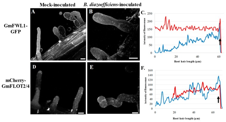
Figure 4.
Subcellular localization of GmFWL1-GFP (A–C) and mCherry-GmFLOT2/4 (D,E) in mock- and B. diazoefficiens-inoculated soybean root hairs in response to cytokinin treatment. C and F: Quantification of the intensity of the fluorescence (y-axis) of the GmFWL1-GFP (C) and mCherry-GmFLOT2/4 proteins (F) in mock- (red) and B. diazoefficiens-inoculated (blue) and cytokinin-treated soybean root hair cells (x-axis, μm; the black arrow highlight the position of the root hair tip). Scale = 10 μm. The pictures shown are representative from a series of observations conducted on three independent biological experiments.

