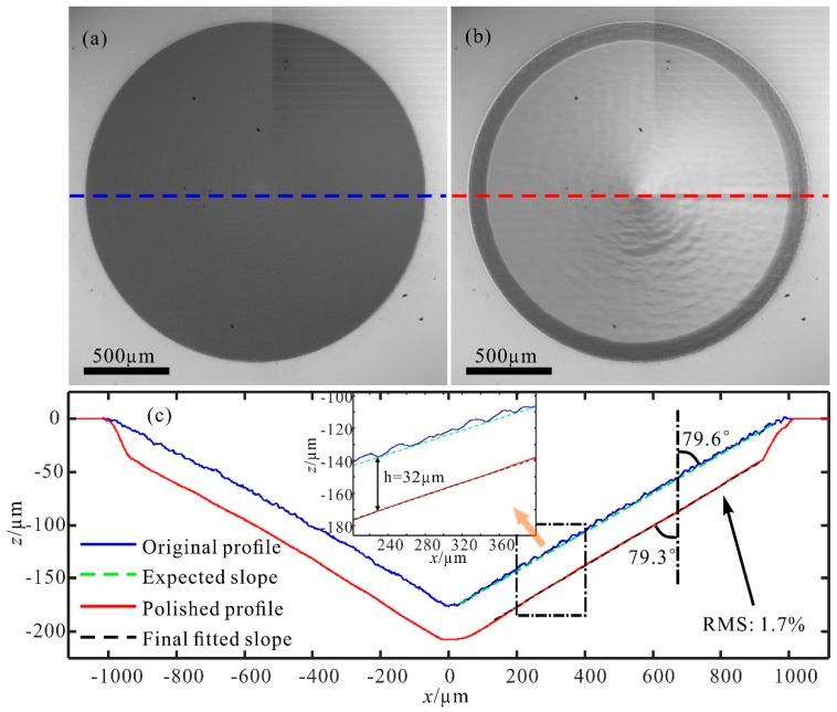
Figure 6.
Optical microscope images of (a) the original multi-stage structure and (b) the structure after laser ablation. (c) Cross-section views of the multi-stage structure before and after polishing. The insert is an enlarged view of the area in the rectangle. Profiles are characterized by a stylus profiler with a vertical (z-direction) resolution of 8 nm and a transversal (x-direction) resolution of 0.1 µm. Slopes (dashed lines) were fitted according to the profiles.

