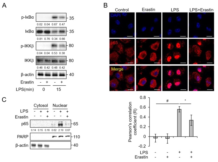
Figure 6.
Erastin blocks LPS-induced NF-κB activation in bone marrow-derived macrophages (BMDMs). (A) Phosphorylation of IκBα and IKKβ in BMDMs was examined using western blot analysis. BMDMs were pre-incubated with DMSO or erastin in DMSO (20 μM) for 2 h and subsequently inducted with 1 μg/mL of LPS for the indicated times; β-actin was used as a loading control. The numbers below each band indicate quantitative analysis of target protein relative to β-actin. (B) Nuclear translocation of NF-κB p65 subunit was detected by immunofluorescence staining in BMDMs after pre-treatment with or without 20 μM of erastin for 2 h, then induction with medium containing 1 μg/mL of LPS for 30 min. Scale bar, 10 μm. Graph represents the co-localization of p65 and DAPI by Pearson’s correlation coefficient. Number of images = 5. (C) Western blot analysis of p65 in the cytosol and nuclear fraction of BMDMs with or without pre-incubation with erastin (20 μM), followed by activation with 1 μg/mL of LPS for 30 min; PARP and β-actin were used as nuclear and cytosolic fraction loading controls. The numbers below each band indicate quantitative analysis of target protein relative to β-actin and PARP. Statistical analyses were performed using paired two-tailed Student’s t-test. * p < 0.05, # p < 0.01.

