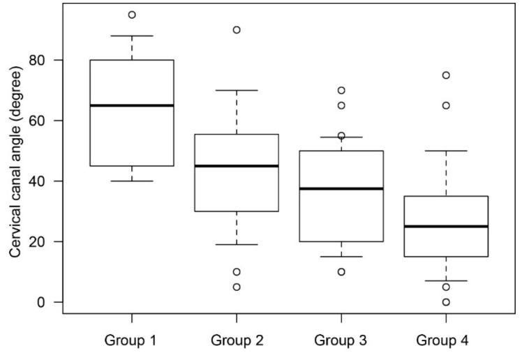
Figure 3.
The mean cervical canal angle in cases without posterior extrauterine wall adhesion. The cervical canal angle was analyzed to evaluate physiological changes in the cervical canal according to gestational age at MRI. In this analysis, cases with posterior adhesion were excluded. The vertical axis presents the cervical canal angle and the horizontal axis presents the patient groups divided according to gestational age on MRI. Mean cervical canal angles were 64.4° in group 1, 42.8° in group 2, 36.4° in group 3, and 28.4° in group 4.

