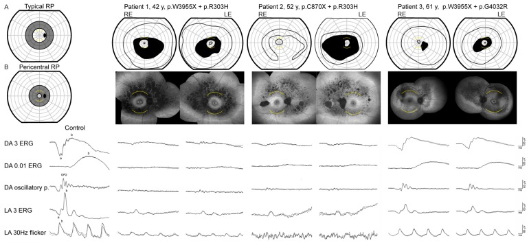
Figure 1.
Goldmann visual field (first row) fundus autofluorescence (second row) and full-field ERG (bottom row) of three USH2A patients with double hyperautofluorescent rings. Goldmann perimetry was performed using II/1 and II/4 targets. The arcuate yellow lines mark the 15° radius on Goldmann visual fields and FAF images. RE = right eye, LE = left eye. The figures (A) and (B) mark the approximate location of scotoma associated with “typical” and “pericentral” RP according to Sandberg et al. [9]. Patients 1 and 2 had absent rod responses and residual cone responses in keeping with the diagnosis of RP. Patient 3 had a mild reduction of the rod (DA 0.01 ERG) and cone (LA 3 ERG and LA 30 Hz flicker) specific responses in a cone-rod dysfunction pattern. The first column shows ERG responses of a representative healthy control.

