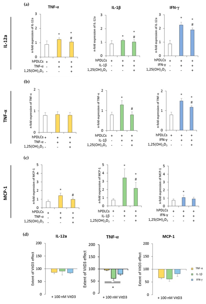
Figure 2.
The expression of pro-inflammatory genes in in vitro differentiated CD68+ macropahges after coculture with hPDLSCs primed with 1,25(OH)2D3 and different cytokines. Primary hPDLSCs were primed with 10 ng/mL TNF-α or 5 ng/mL IL-1β or 100 ng/mL IFN-γ in the absence or presence of 100 nM 1,25(OH)2D3. In vitro differentiated CD68+ macrophages were applied to the indirect coculture system with primed hPDLSCs. Gene expression levels of IL-12a (a), TNF-α (b), and monocyte chemoattractant protein (MCP)-1 (c) were determined in macrophages using quantitative polymerase chain reaction (qPCR) after 24 h of coculture. (a–c) shows the n-fold expression of indicated pro-inflammatory cytokines. In vitro differentiated CD68+ macrophages without hPDLSCs served as control (n-fold expression = 1). (d) Shows the extent of the effect of 1,25(OH)2D3 on the macrophage functional status regarding the presence of differently-primed hPDLSCs (expressed in % of corresponding cytokine treatment). All data are presented as mean ± S.E.M. from five independent experiments with hPDLSCs isolated from five different patients. (a–c): * significantly different (p < 0.05) compared to macrophages in the presence of unprimed hPDLSCs. # Significantly different (p < 0.05) compared between macrophages in the presence of primed hPDLSCs with and without 1,25(OH)2D3. (d): * significantly different (p < 0.05) compared between groups as indicated.

