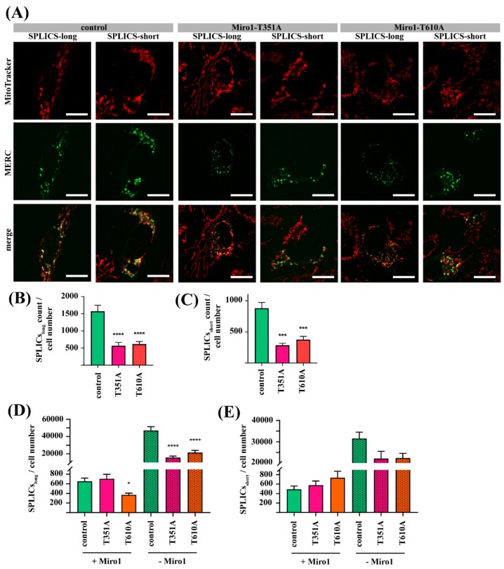
Figure 5.
(A) Immortalized fibroblasts were transfected either with the SPLICS-long, or with the SPLICS-short construct. After 12 h, cells were stained with MitoTracker deep red FM for live cell imaging, using a 63x objective; scale bars indicate 20 µm. (B) Quantification of wide MERCs (from SPLICS-long signal) and (C) narrow MERCs (from SPLICS-short signal) per cell. Fibroblasts transfected with (D) SPLICS-long or (E) SPLICS-short constructs were fixed and stained with an antibody against Miro1. Afterwards, cells were imaged with a 63× objective and SPLICS signals with and without Miro1 were quantified. All data indicated as mean ± SEM. Significance was assessed using a Kruskal-Wallis test (n = 3; ~16 cells analyzed per fibroblast line per experiment). * p < 0.05; *** p < 0.0001; **** p < 0.00001.

