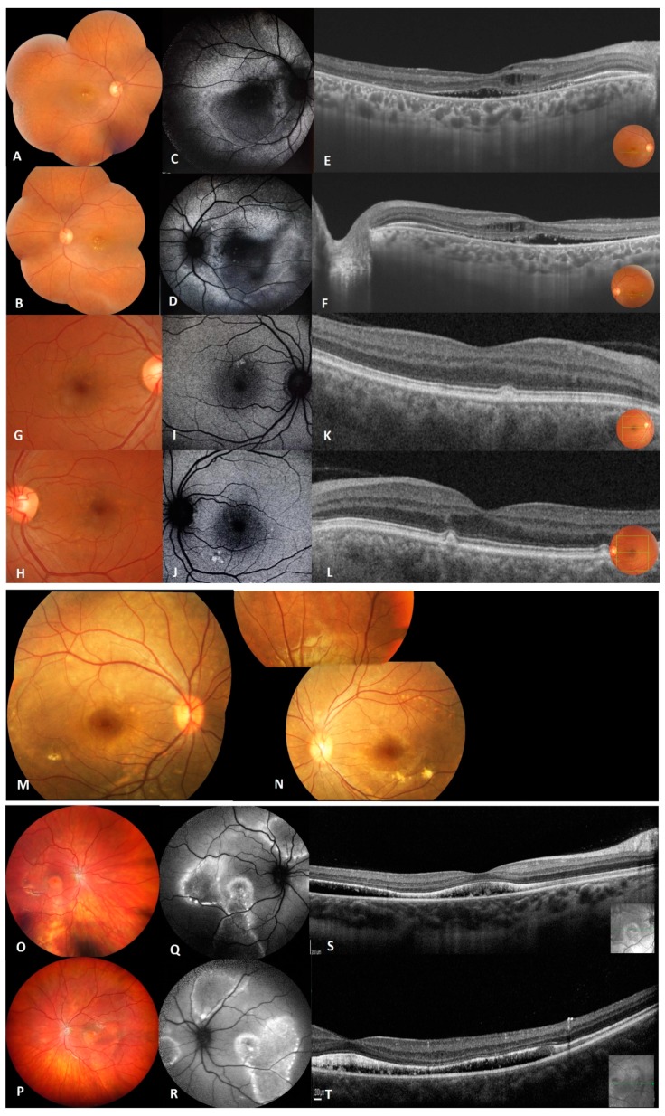
Figure 2.
Clinical and imaging features of patients from family A (A–L), family B (M,N) and family C (O–T). (A,B) Fundus photographs of right and left eye of proband (II.1) of family A showing macular vitelliform lesions with yellow flecks and dots extending to the mid-periphery. (C,D) Fundus autofluorescence images of proband showing macular hypo-FAF surrounded by marked increased autofluorescence. (E,F) Macular OCT images of the proband showing hyperreflective accumulations on RPE, cystoid intra-retinal and serous subretinal fluid. (G–J) Fundus photographs and FAF of the sibling II.2 showing macular yellowish autofluorescent deposits. (K,L) OCT of both eyes of patient II.2 with very small focal subretinal macular deposits.(M,N) Fundus photographs of right and left eye of proband (II.3) from family B showing focal areas of sparse vitelliform deposits in the posterior pole. (O,P) Fundus photographs of right and left eye of propositus (II.1) from family C showing multiple yellowish, vitelliform deposits in the macula and along the vessel arcades with yellowish deposits on the optic disc. (Q,R) Fundus autofluorescence images of proband showing hyper autofluorescence zones along the vascular arcades and in the posterior pole. (S,T) OCT of the proband’s macula revealing bilateral diffuse flat serous subretinal fluid.

