Figure 3.
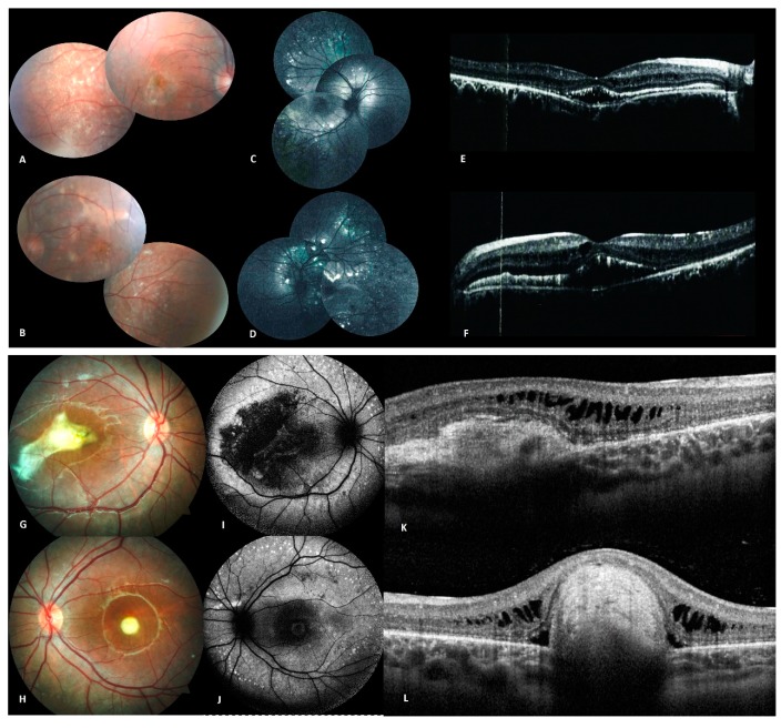
Clinical and imaging features of patients from family D (A–F), and family E (G–L). (A,B) Fundus photographs of both eyes of propositus (II.1) from family D showing multiple yellowish round vitelliform lesions, as well as subretinal fibrosis. (C,D) Autofluorescence image of both eyes showing hyper autofluorescent dots in the posterior pole and along the vascular arcades. (E,F) OCT with hyperreflective macular subretinal deposits with intra-retinal cysts. (G,H) Fundus photography of both eyes of propositus (II.1) from family E showing a macular yellow lesion with fibrosis in the right eye and a macular yellow round lesion in the left eye. (I,J) Autofluorescence image of both eyes with autofluorescent dots scattered along the vascular arcades. (K,L) OCT showing subretinal deposits with intraretinal fluid in the right eye and a subretinal mass in the left eye.

