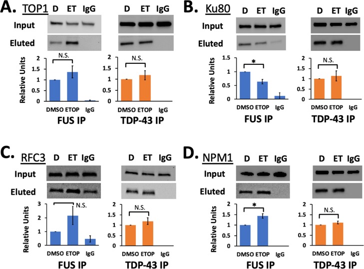
Figure 4.
TDP-43 binds DNA damage repair proteins. co-IP was performed for LAP-tagged FUS (blue) and TDP-43 (orange). Western blots show inputs and eluted proteins for TOP1 (A), Ku80 (B), RFC3 (C), and NPM1 (D). Levels of protein in western analyses were quantified and then normalized to western blots for the input samples and for the eluted LAP-FUS or LAP-TDP43. Only changes for LAP-FUS interacting with Ku80 and NPM1 were significant (p < 0.05, Student’s t-test assuming equal variances). Each western blot includes samples treated with DMSO (D), 5 μM etoposide for 1 h (ET), and pulldown with a negative control nonspecific IgG antibody (IgG). Error bars show standard error from three or four biological replicates.

