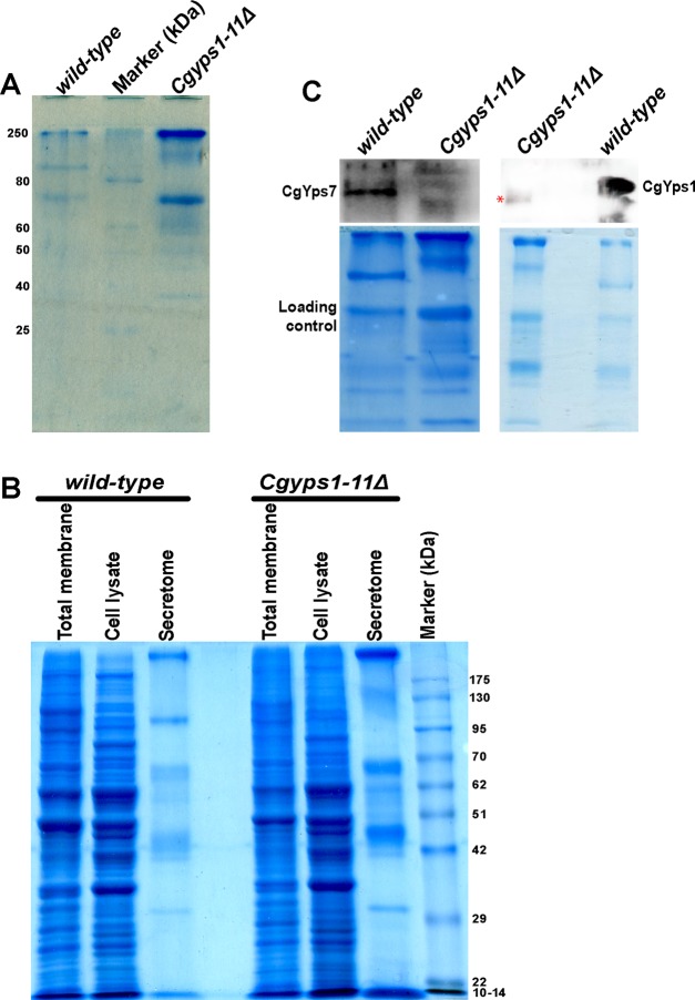
Figure 8.
CgYps1 and CgYps7 are secreted into the medium. (A) Representative SDS-PAGE gel image indicating increased protein secretion into the medium of the Cgyps1-11Δ mutant. The secretomes of wild-type and Cgyps1-11Δ mutants were collected after 11 doublings in the YNB medium, and 50 μL were resolved on a 12% SDS-PAGE gel. Proteins were stained with Coomassie Brilliant Blue (CBB) for visualization. (B) Representative SDS-PAGE gel image depicting the total membrane, cell lysate, and secretory protein profiles of wild-type and Cgyps1-11Δ mutants. Equal volume of secretomes (50 μL) and 100 μg of total membrane and cell lysate were resolved on a 10% SDS-PAGE gel and stained with CBB for visualization. (C) Representative western blot images of CgYps1 and CgYps7 indicating their secretion into the medium of the wild-type strain. Equal volume (50 μL) of secretomes of wild-type and Cgyps1-11Δ strains were loaded on a 10% SDS-PAGE and resolved for 4 h. Proteins were transferred to the polyvinylidene fluoride membrane and probed with anti-CgYps1 and anti-CgYps7 antibodies. CBB-stained SDS-PAGE gels were used as loading control. Of note, the red asterisk marks a nonspecific band seen in the Cgyps1-11Δ secretome.

