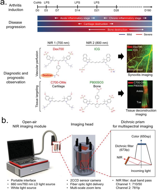
Figure 1.

Overall strategy of imaging for the diagnosis and prognosis of RA. a) Preparation of a CAIA mouse model and dual‐channel NIR imaging along with RA disease progression (top), and chemical structures of the NIR fluorophores with the following outcome from an in vivo assessment (bottom). Red and lime green colors represent 700 and 800 nm NIR, respectively. b) Open‐air, intraoperative optical imaging system equipped three excitation light sources, a 2CCD camera, and multi‐scale zoom lens. Details of imaging head and dichroic prism‐based beam filtration for visible and NIR lights are described in the schematic drawing.
