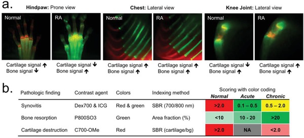
Figure 5.

Real‐time dual‐channel intraoperative imaging of cartilage and bone. a) 25 nmol (0.05 mg kg−1) of C700‐OMe and P800SO3 was injected into the same mice with CAIA 2 h and 24 h prior to imaging, respectively. Cartilage and bone of central skeleton in the chest area, large joint in the knee area, and peripheral small joints in the hindpaw area were observed in the same RA model. Red and lime green pseudocolors were used for 700 and 800 nm NIR, respectively, in the color‐NIR merged images. b) Permissive scoring index for the prognosis of RA. The metric was based on the SBR and area fraction (%) of synovial inflammation, bone resorption, and cartilage destruction, and the color codes were provided for a convenient visual interpretation of inflammatory severity and disease progression.
