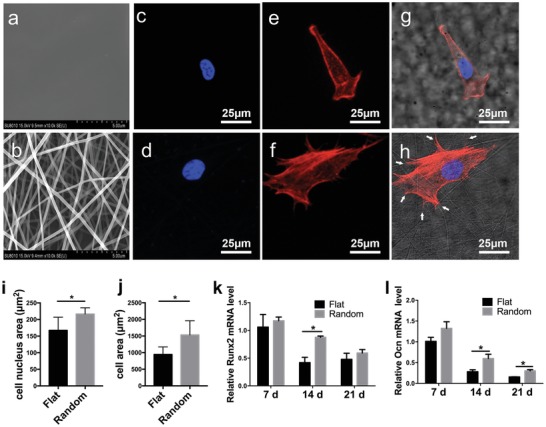
Figure 1.

Morphology and osteogenic differentiation of human bone marrow‐derived stem cells (hBMSCs) on poly‐l‐lactide (PLLA) fibrous scaffolds and cast PLLA films. a) Representative scanning electron microscopy images showing the smooth surface in the flat group. b) Representative scanning electron microscopy image of randomly arranged nanofibers (scale bar, 5 µm). c,d) Confocal immunofluorescence staining of nuclei with 4′,6‐diamidino‐2‐phenylindole (DAPI) in hBMSCs from the c) flat group and d) the random fiber group after 4 h in culture. e,f) Confocal immunofluorescence staining of F‐actin with rhodamine‐labeled phalloidin in hBMSCs from the e) flat group and f) the random fiber group after 4 h in culture. g) Merged confocal images of (c) and (e), with bright‐field microscopy images of the flat group. h) Merged confocal microscopy images of (d) and (f), with bright‐field microscopy images of the random group. Scale bar, 25 µm. Quantification of i) the nuclear size and j) cell spreading area of hBMSCs in the flat group and random group (n = 30). j) Quantification of hBMSCs in the flat and random groups (n = 30). k) Runx2 and l) Ocn mRNA levels in hBMSCs in the flat and random groups at 7, 14, and 21 days, respectively. Results are means ± SEM (n = 3). *p < 0.05, * by two‐sample t‐test.
