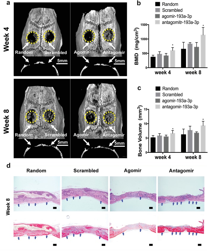
Figure 4.

Nanofiber membranes loaded with miR‐193a‐3p antagomir enhanced the healing of critical‐sized bone defects. a) Representative micro‐CT images and sagittal views of rat cranial critical‐sized full‐thickness defects at 4 and 8 weeks after surgery (scale bar, 5 mm). Yellow circles and white arrows indicate the bone defect area. b,c) Quantitative analysis of BV and BMD of the newly formed bone. Data are means ± SE) (n = 6) and all p‐values are based on one‐way ANOVA with a post hoc test (*p < 0.05). d) Histological results of 8‐weeks H&E staining (Top row) and Masson staining (Bottom row). Blue arrows denote the newly‐formed bone. (bar = 200 µm)
