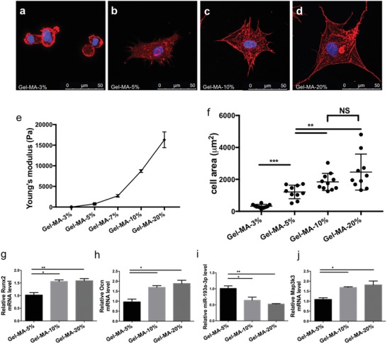
Figure 8.

miR‐193a‐3p mediated hBMSC osteogenic differentiation in response to substrate stiffness on gelatin methacrylate (Gel‐MA). a–d) Confocal immunofluorescence of cytoskeletal actin and nuclear DNA of hBMSCs cultured on Gel‐MA with different substrate stiffnesses at 24 h postseeding (scale bar, 50 µm). Actin, red; nuclear DNA, blue. e) The stiffness of the Gel‐MA hydrogel could be modified by altering its concentration; the higher the gel concentration, the higher the Young's modulus. f) Cell spreading areas on substrates with different stiffnesses (n = 10). g–j) mRNA levels of Runx2, Ocn, miR‐193a‐3p, and Map3k3 in cells cultured on substrates with different stiffnesses (n = 3). Results are means ± SEM. Samples were subjected to one‐way ANOVA with Tukey's post hoc test. Significant differences are denoted by *p < 0.05.
