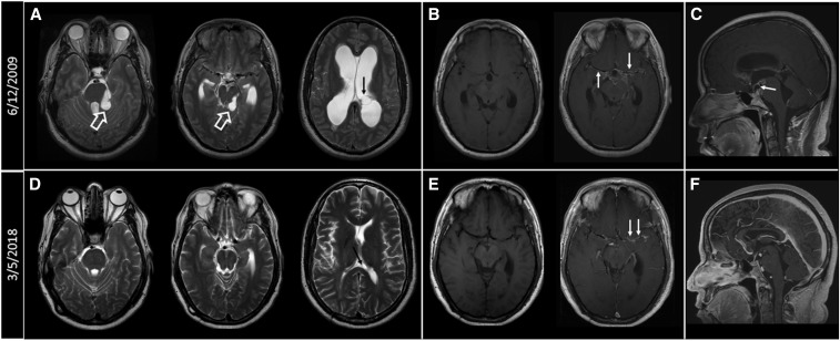
Figure 2.
(A) Axial T2-weighted images at the levels of the pons, midbrain, and lateral ventricles. Cystic masses are seen in the perimesencephalic cistern (white hollow arrows) with secondary distortion of adjacent brain stem structures. Hydrocephalus is noted with transependymal cerebrospinal fluid seepage. A left lateral ventricular cyst can also be seen (black arrow). (B) Pre- and postcontrast T1-weighted images showing abnormal enhancement along the courses of the middle cerebral arteries (MCAs) and anterior cerebral arteries (ACAs) and extending into the interpeduncular cistern (white arrows). (C) Sagittal postcontrast T1-weighted images showing suprasellar rim enhancing cyst anteriorly displacing the pituitary stalk (white arrow). (D) Axial T2-weighted images obtained on March 5, 2018, showing resolution of cystic masses and mass effect on the brain stem structures. The ventricular cyst has markedly decreased in size (black arrow). (E) Pre- and postcontrast T1-weighted images show significant improvement of the perivascular and meningeal enhancing lesions (white arrows). (F) Sagittal postcontrast T1-weighted images show resolution of the suprasellar cyst.

