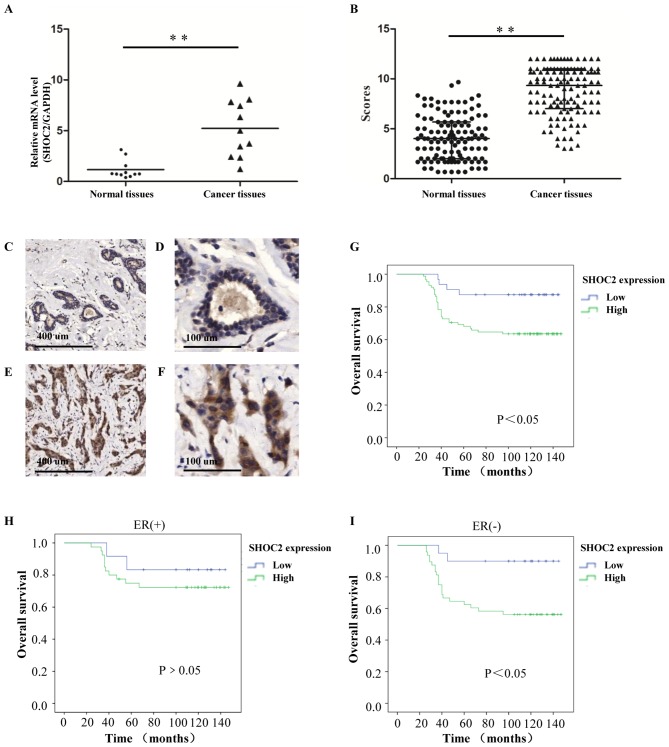
Figure 3.
Clinical association and prognostic value of SHOC2 expression for breast cancer. (A) Relative mRNA expression levels of SHOC2 in fresh clinical samples. (B) IHC staining scores for the normal breast and invasive breast cancer tissues. (C and D) IHC staining shows that SHOC2 expression was low in normal breast tissues; (C) magnification, ×50 and (D) magnification, ×200. (E and F) IHC staining shows that SHOC2 was upregulated in invasive breast cancer tissues; (E) magnification, ×50 and (F) magnification, ×200. (G) Kaplan-Meier plot showing a significant association between SHOC2 expression and OS for patients with breast cancer. (H) In the ER (+) subgroups, OS rates were not significantly affected by SHOC2 expression. (I) In the ER (−) subgroups, OS rates were significantly affected by SHOC2 expression. **P<0.01. ER, estrogen receptor; IHC, immunohistochemistry; OS, overall survival; SHOC2, SHOC2 leucine rich repeat scaffold protein.

